Forty-third ISSUE
September 20, 2023
Changes in health care costs, survival, and time toxicity in the era of immunotherapy and targeted systemic therapy for melanoma
JAMA Dermatology
JAMA Dermatology

Chemo? How about chem-no!
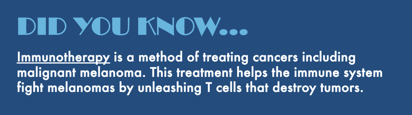
The advent of modern immunotherapies and targeted therapies for melanoma has caused melanoma treatment to evolve over the last decade. However, the impact these therapies have on both outcomes and costs remains unclear. This retrospective cohort study compared a propensity matched cohort of 731 patients with stage II to IV melanoma from January 2018 - March 2019 and 731 patients with invasive melanoma from January 2007 - December 2012.
What did they find?
Limitations: This study did not assess long-term costs associated with ongoing treatment, recurrence, or surveillance using modern therapies. Time toxicity did not include virtual visits, and thus may underestimate the true total time burden experienced by patients.
Main Takeaway: Patients treated with modern immunotherapy and targeted therapy for melanoma have increased overall survival, but place a higher economic burden on the healthcare system.
The advent of modern immunotherapies and targeted therapies for melanoma has caused melanoma treatment to evolve over the last decade. However, the impact these therapies have on both outcomes and costs remains unclear. This retrospective cohort study compared a propensity matched cohort of 731 patients with stage II to IV melanoma from January 2018 - March 2019 and 731 patients with invasive melanoma from January 2007 - December 2012.
What did they find?
- The 2018 - 2019 cohort had higher mean health care costs ($47,886 +/- $55,176 versus $33,347 +/- $31,576, standard difference 0.32)
- This was especially true for patients with stage III ($67,108 +/- $57,226 versus $46,511 +/- $30,622) and stage IV disease ($117,450 +/- $79,272 versus $47,739 +/- $37,652)
- The 2018 - 2019 cohort had a greater overall 3-year survival
- 74.2% [95% CI, 70.8%-77.2%] versus 65.8% [95% CI, 62.2%-69.1%], hazard ratio 0.72 [95% CI, 0.61-0.85]; p<0.001
- Overall, time toxicity (i.e., time spent in contact with healthcare system) was similar between the two time periods
Limitations: This study did not assess long-term costs associated with ongoing treatment, recurrence, or surveillance using modern therapies. Time toxicity did not include virtual visits, and thus may underestimate the true total time burden experienced by patients.
Main Takeaway: Patients treated with modern immunotherapy and targeted therapy for melanoma have increased overall survival, but place a higher economic burden on the healthcare system.
Cross-sectional study shows Asian American and Pacific Islanders with melanoma have increased odds of treatment delays
Journal of the American Academy of Dermatology
Journal of the American Academy of Dermatology
Melanoma Treatment: Let’s Outrun the Sun…and the Clock!
Cancer is the leading cause of death among Asian Americans. Asian Americans and Pacific Islanders (AAPI) have a 27% higher risk of mortality from melanoma than non-Hispanic White (NHW) patients.
This cross-sectional study utilized the National Cancer Database to examine differences in time from diagnosis to definitive surgery (TTDS) between AAPI patients and NHW patients and explore variables that may contribute to a potential difference.
What did they find:
Main takeaway: Despite living statistically closer to hospitals and having a higher average income, AAPI patients with melanoma experienced a longer TTDS than their NHW counterparts, highlighting a disparity in melanoma treatment.
Limitations: AAPI patients comprised 0.33% of the sample
Cancer is the leading cause of death among Asian Americans. Asian Americans and Pacific Islanders (AAPI) have a 27% higher risk of mortality from melanoma than non-Hispanic White (NHW) patients.
This cross-sectional study utilized the National Cancer Database to examine differences in time from diagnosis to definitive surgery (TTDS) between AAPI patients and NHW patients and explore variables that may contribute to a potential difference.
What did they find:
- AAPI patients had an average TTDS of 44 days compared to 37 days for NHW patients (p < 0.001)
- AAPI patients had 1.5 times the odds of a TTDS between 61 and 90 days (95% CI, 1.26-1.87; p < 0.001) and twice the odds of a TTDS >90 day (95% CI, 1.59-2.58; p < 0.001)
- AAPI melanoma patients often presented with later-stage melanoma (p < 0.001)
- Racial differences in TTDS persisted amongst those with Medicare and private insurance
- On average, AAPI patients lived closer to the hospital than NHW (78% vs 63% less than 20 miles from the hospital; p < 0.001)
- AAPI patients were more likely to be in the top quartile for median household income (63% vs 52%), while NHW patients were more likely to be in the lower 3 quartiles (p < 0.001)
Main takeaway: Despite living statistically closer to hospitals and having a higher average income, AAPI patients with melanoma experienced a longer TTDS than their NHW counterparts, highlighting a disparity in melanoma treatment.
Limitations: AAPI patients comprised 0.33% of the sample
Dermatofibroma versus dermatofibrosarcoma protuberans: a nuclear morphology study
American Journal of Dermatopathology
American Journal of Dermatopathology

Keep an eye on the nuclei
Dermatofibrosarcoma protuberans (DFSP) is a locally invasive soft tissue sarcoma that shares histologic similarities with the common, benign dermatofibroma (DF). DFSP can be distinguished from DF using immunohistological stains for clusters of differentiation 34 (CD34) and Factor XIIIa, however, these stains are expensive and neither sensitive nor specific.
Histologically, DFSP nuclei have an alternating ovoid and narrow spindle shape throughout the tumor sample. Researchers were curious if this nuclear morphology can be used to distinguish DFSP from DF. Histologic slides of 246 DF and 85 DFSP samples were reviewed retrospectively and evaluated for the presence or absence of alternating ovoid-spindled nuclear morphology.
What did they find?
Limitations: All histologic slides were interpreted by a single reviewer which may have influenced the results of the study.
Main takeaways: Predominant alternating ovoid-spindled nuclear morphology is highly sensitive and specific for DFSP, and can be used as a cost-effective diagnostic tool to distinguish DFSP from DF.
Dermatofibrosarcoma protuberans (DFSP) is a locally invasive soft tissue sarcoma that shares histologic similarities with the common, benign dermatofibroma (DF). DFSP can be distinguished from DF using immunohistological stains for clusters of differentiation 34 (CD34) and Factor XIIIa, however, these stains are expensive and neither sensitive nor specific.
Histologically, DFSP nuclei have an alternating ovoid and narrow spindle shape throughout the tumor sample. Researchers were curious if this nuclear morphology can be used to distinguish DFSP from DF. Histologic slides of 246 DF and 85 DFSP samples were reviewed retrospectively and evaluated for the presence or absence of alternating ovoid-spindled nuclear morphology.
What did they find?
- 80 of 82 (97.6%) DFSP samples showed predominant alternating ovoid-spindled nuclear morphology compared to 0 of 242 DF samples (P<0.001)
- Alternating ovoid-spindled nuclear morphology was found to be highly specific (Sp=1) for DFSP
- Alternating ovoid-spindled nuclear morphology was found to be highly sensitive (Sn=0.98) for DFSP
Limitations: All histologic slides were interpreted by a single reviewer which may have influenced the results of the study.
Main takeaways: Predominant alternating ovoid-spindled nuclear morphology is highly sensitive and specific for DFSP, and can be used as a cost-effective diagnostic tool to distinguish DFSP from DF.
Intralesional vitamin D3, CO2 laser, and combined vitamin D3 and CO2 laser: what is the best treatment for palmoplantar warts?
Dermatologic Surgery
Dermatologic Surgery
Don’t be a worry wart!
Palmoplantar warts, caused by the human papillomavirus, are common. There are currently no treatments which provide complete response or prevent recurrence. CO2 laser therapy is one treatment that vaporizes and excises the affected area, while treatment with intralesional vitamin D3 immunotherapy likely helps regulate cell proliferation and appropriate immune system activation.
In this study, the efficacy of treating palmoplantar warts with a combined treatment of vitamin D3 injection and CO2 laser was compared to the efficacy of either treatment on its own. 80 patients were randomly assigned into 4 treatment groups: Vitamin D3 injection, CO2 laser, combined vitamin D3 injection followed by CO2 laser followed by 3 more D3 injections, and normal saline injection acting as the control group. Outcomes were measured by physician assessment, photography, and patient satisfaction.
What did they find?
Main Takeaways: Palmoplantar warts can be effectively treated with CO2 laser, intralesional vitamin D3, and combined vitamin D3 and CO2 laser; however, there is no significant difference in response to treatment or recurrence. CO2 laser may not be the treatment of choice for diabetics or older aged individuals due to slow wound healing.
Palmoplantar warts, caused by the human papillomavirus, are common. There are currently no treatments which provide complete response or prevent recurrence. CO2 laser therapy is one treatment that vaporizes and excises the affected area, while treatment with intralesional vitamin D3 immunotherapy likely helps regulate cell proliferation and appropriate immune system activation.
In this study, the efficacy of treating palmoplantar warts with a combined treatment of vitamin D3 injection and CO2 laser was compared to the efficacy of either treatment on its own. 80 patients were randomly assigned into 4 treatment groups: Vitamin D3 injection, CO2 laser, combined vitamin D3 injection followed by CO2 laser followed by 3 more D3 injections, and normal saline injection acting as the control group. Outcomes were measured by physician assessment, photography, and patient satisfaction.
What did they find?
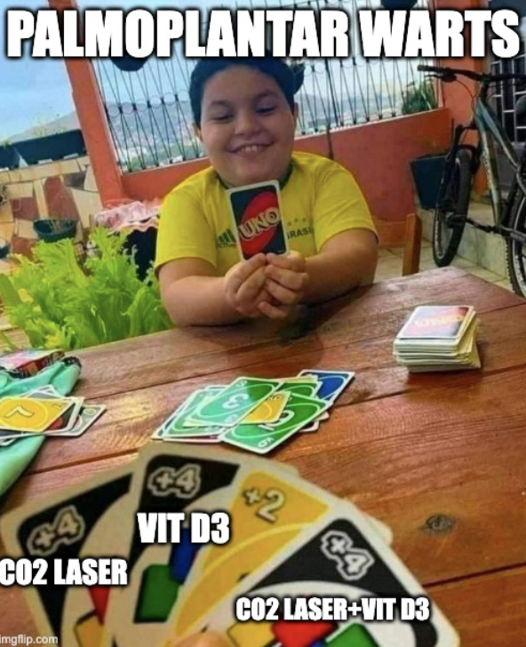
- The Vit D, CO2 laser, and combined treatment groups showed a complete clearance in 80%, 75%, and 90% of their patients respectively
- There was no statistically significant difference in response or recurrence between the 3 intervention groups, but there was a significant difference between the 3 intervention groups vs. control
- There was a negative correlation between the degree of response and age in the CO2 laser and combined group (r = -0.492, p = .028; r = -0.451, p = .004)
- Significant delayed onset of response was found in patients in the older than 40 (8.86 weeks) age group compared to the <20 (6.67 weeks) and 20-40 (6.80 weeks) age groups (p = .019) in all treatment groups
- Significantly fewer side effects were seen in the laser only group compared to the other two interventions (0% vs 40% and 45%, p = .000); side effects in the vitamin D and combined treatment had a significant negative correlation with age
Main Takeaways: Palmoplantar warts can be effectively treated with CO2 laser, intralesional vitamin D3, and combined vitamin D3 and CO2 laser; however, there is no significant difference in response to treatment or recurrence. CO2 laser may not be the treatment of choice for diabetics or older aged individuals due to slow wound healing.
What is the safety and efficacy of PDT-activated aminolevulinic acid 20% solution for the treatment of facial cutaneous SCCis?
Journal of Cosmetic Dermatology
Journal of Cosmetic Dermatology

A new way to treat SCCis….one laser at a time!
Squamous Cell Carcinoma (SCC) is the second most common cutaneous cancer. Mohs surgery is the gold standard for therapy, though nonsurgical treatment, including photodynamic therapy (PDT), is also widely used. Researchers aimed to assess the safety and efficacy of PDT-activated aminolevulinic acid (ALA) 20% topical solution for treatment of facial cutaneous SCC in situ (SCCis).
This prospective clinical trial included patients (n=20) with biopsy-confirmed untreated facial SCCis. Topical ALA 20% solution was applied to the lesion and adjacent skin followed by pulsed dye laser (0.45 ms at 13 J/cm2) and blue light illumination (16 min at 10 J/cm2) 24 hours later. A second ALA-PDL-PDT treatment was administered after 30 days. Surgical excision was performed 4-6 weeks post treatment. Investigators assessed clearance, pigmentation, tolerability, and skin reactions at each visit.
What did they find:
Main takeaway: Aminolevulinic acid 20% solution with PDL-PDT activation is a safe and well-tolerated treatment option for facial squamous cell carcinoma in situ.
Squamous Cell Carcinoma (SCC) is the second most common cutaneous cancer. Mohs surgery is the gold standard for therapy, though nonsurgical treatment, including photodynamic therapy (PDT), is also widely used. Researchers aimed to assess the safety and efficacy of PDT-activated aminolevulinic acid (ALA) 20% topical solution for treatment of facial cutaneous SCC in situ (SCCis).
This prospective clinical trial included patients (n=20) with biopsy-confirmed untreated facial SCCis. Topical ALA 20% solution was applied to the lesion and adjacent skin followed by pulsed dye laser (0.45 ms at 13 J/cm2) and blue light illumination (16 min at 10 J/cm2) 24 hours later. A second ALA-PDL-PDT treatment was administered after 30 days. Surgical excision was performed 4-6 weeks post treatment. Investigators assessed clearance, pigmentation, tolerability, and skin reactions at each visit.
What did they find:
- Excluding those with skip lesions, 94% (n=17) of patients achieved histological clearance at the end of treatment and excision
- 65% (n=13) of patients had observed clinical clearance prior to surgical excision
- The average pain score recorded by patients after each PDT administration was 2.95
- 65% (n=13) of patients did not report adverse events. Those that did were mostly allergic contact dermatitis at the treatment site
Main takeaway: Aminolevulinic acid 20% solution with PDL-PDT activation is a safe and well-tolerated treatment option for facial squamous cell carcinoma in situ.

We live in an era where “physical perfection” is celebrated on social media and the demand for non-surgical cosmetic procedures continues to surge. This article investigates the positive and negative effects of cosmetic procedures on patient mental and emotional well-being. We reviewed articles discussing the psychological effects of cosmetic dermatologic procedures.
What we found:
It is crucial that physicians be aware of the psychological consequences of cosmetic procedures. Open communication during consultations is crucial to address psychological risks and promote holistic patient care.
What we found:
- Cosmetic procedures have positive and negative psychological impacts
- Image-altering filters on social media have influenced individuals' perceptions of appearance
- Post-pandemic anxieties contributed to increased interest in cosmetic alterations
- Patients with body dysmorphic disorder (BDD) may be at higher risk for dissatisfaction after cosmetic procedures
It is crucial that physicians be aware of the psychological consequences of cosmetic procedures. Open communication during consultations is crucial to address psychological risks and promote holistic patient care.