third issue
february 9, 2022
How are outcomes different in patients after Hidradenitis supprativa (HS) surgery?
JAMA Dermatology
HS surgery? Yes, recommend. Prior studies have shown that deroofing and excision for HS have led to positive outcomes, but have not considered the patient experience with post-op. Furthermore, most of these have featured white individuals, yet HS is significantly more predominant in Black individuals. This retrospective cohort study analyzed survey responses from predominantly Black patients who had HS surgery from April 2014 to December 2018 (n = 78). The study was limited to procedures performed by a single surgeon, as well as by potential recall bias. However, results showed that longer healing time was significantly associated with non-White race and ethnicity (11.4 days; β, 1.14; P = .02) compared with White race and ethnicity and with local excision with closure (0.9 days; β, 0.08; P = .003) compared with deroofing. Although diabetes, smoking, and morbid obesity are typically considered risk factors for poor healing, these variables were not significantly associated with recovery metrics in the present sample. Therefore patients with these comorbidities should be aware of the risks, but it should not be a contraindication. Furthermore, dermatologists should consider their patients’ demographics and subsequently provide more accurate expectations about recovery post-op before recommended HS surgery.
Is Mohs for intermediate-risk squamous cell carcinoma more cost effective than wide local excision?
Journal of American Academy of Dermatology
That feeling when you thought you were overpaying for something but then it turns out to be worth it! While Mohs micrographic surgery (MMS) has higher cure rates for treatment of cutaneous squamous cell carcinoma (cSCC) as compared with wide local excision (WLE), critics have cited its cost as a downside. However, recent studies have only analyzed upfront costs and have not calculated “cost effectiveness”. This cost-effectiveness analysis conducted at Brigham and Women’s Hospital evaluates cost effectiveness of MMS v WLE for T2a cSCC over a 5-year time period. The authors found that MMS was $333.83 less expensive than WLE ($4365.57 [95% CI, $3664.68-$6901.66] vs $4699.41 [95% CI, $3782.94-$10,019.31]). MMS gained 3.733 quality-adjusted life years (QALY) over 5 years. Threshold analyses showed that even if WLE costs were equal to $0, MMS would still be preferred because of the increased QALY over WLE. Based on this analysis, it is safe to say that MMS is less costly and more effective than WLE. However, this is only a single study and was not randomized. Future studies should be conducted in higher risk tumors.
Debugging with Dupilumab
Pediatric Dermatology
Dupilumab is the hot new medication for moderate-to-severe atopic dermatitis (AD). In October dupilumab received FDA approval for children in the 6-11 age group. With AD often presenting in childhood, this approval can be life changing for those who qualify for treatment. Children with AD are at increased risk of contracting infections because of skin barrier dysfunction and immunosuppression from traditional AD medications. Dupilumab differs from traditional immunosuppressive medications because it targets IL4 and IL13 - cytokines not believed to play a major role in host-defense. This study investigated how dupilumab therapy influenced infection risk. Data was pooled from two 16-week, randomized-controlled trials of adolescents ages 12-17. 612 participants were included in the analysis, 205 of which were placebo and 407 of whom received dupilumab. Analysis showed the total number of skin infections were significantly lower in those treated with dupilumab compared to control (p=0.001). Additionally, the number of total infections was numerically decreased and approached statistical significance (p=0.051). One limitation of this study is that diagnoses were clinically made and did not always have microbiological confirmation. Overall, there was a reduced frequency of skin infections in children who were being treated with dupilumab for AD.
QUESTION OF THE WEEK
NEJM CHALLENGE QUESTION
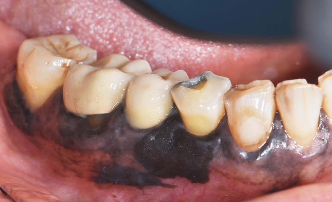
61-year-old woman presented with discoloration along her gums that had rapidly expanded over the past year. What is the diagnosis?
- Amalgam tattoo
- Gingival melanoma
- Kaposi’s sarcoma
- Oral melanoacanthoma
- Physiologic pigmentation
Gingival melanoma
The correct answer is gingival melanoma. Melanomas may arise from the mucosal epithelial lining of the oral cavity and gastrointestinal tract. The patient was referred to the oncology clinic and underwent imaging, which was negative for lymph-node involvement or distant metastases. She subsequently underwent surgical resection of the lesion and had no recurrence at four-month follow up.
Other answers:
A. Amalgam tattoo- The most common cause of oral pigmentation is foreign body tattoos, and typically, these are due to implantation of dental amalgam. These lesions consist of grey, blue or black discoloration, typically on gums of the lower jaw.
C. Kaposi’s sarcoma- Kaposi’s sarcoma presents as red-purplish lesions in the oral cavity, typically in immunocompromised/HIV patients.
D. Oral melanoacanthoma- This is a very rare, benign brown- black pigmented lesion that appears suddenly and can have rapid growth.
E. Physiologic pigmentation- Oral physiologic pigmentation can occur anywhere in the oral cavity, however gingiva is the most common location. It can present as brown to black discoloration and darker skinned individuals are more commonly affected.
