Forty-eighth issue
NOVEMBER 29th, 2023
How does novel immunotherapy for melanoma affect costs, time burden, and survival?
JAMA Dermatology
JAMA Dermatology
We’re in our ‘immunotherapy’ era…
Malignant melanoma (MM) is the fifth most common cancer in the US, representing a significant portion of the $173 billion annual cancer spending. Novel immunotherapies like PD-1 and CTLA-4 inhibitors have become standard treatments for MM. This retrospective cohort study evaluated the cost, time burden, and overall survival (OS) associated with MM immunotherapies.
What did they find?
Limitations: Costs of ongoing treatment, recurrence, surveillance, and individual indirect costs such as cost of transportation and loss of wages were not accounted for in the study. Undisclosed drug discounts were not included. Time burden may be underestimated.
Main Takeaway: Novel immunotherapy may be more expensive and time consuming, but results in better patient survival rates overall.
Malignant melanoma (MM) is the fifth most common cancer in the US, representing a significant portion of the $173 billion annual cancer spending. Novel immunotherapies like PD-1 and CTLA-4 inhibitors have become standard treatments for MM. This retrospective cohort study evaluated the cost, time burden, and overall survival (OS) associated with MM immunotherapies.
What did they find?
- Health care costs:
- Patients had higher average healthcare costs compared to the control
- Mean [SD], treatment $47,886 [$55,176] vs. control $33,347 [$31,576]
- Time burden:
- Similar between cohorts
- Patients with stage IV disease spent >1 day/week in treatment
- Mean [SD], treatment 58.7 [43.8] vs. control 44.2 [26.5] days; p=0.20
- Overall survival (OS):
- OS was greater for treatment compared to control
- 3-year OS: treatment 74.2% [95% CI, 70.8%-77.2%] vs. control 65.8% [95% CI, 62.2%-69.1%], p<0.001
Limitations: Costs of ongoing treatment, recurrence, surveillance, and individual indirect costs such as cost of transportation and loss of wages were not accounted for in the study. Undisclosed drug discounts were not included. Time burden may be underestimated.
Main Takeaway: Novel immunotherapy may be more expensive and time consuming, but results in better patient survival rates overall.
Modified red light 5-aminolevulinic acid photodynamic therapy versus low-dose isotretinoin for moderate-to-severe acne vulgaris
Journal of the American Academy of Dermatology
Journal of the American Academy of Dermatology
Glow up with M-PDT!
Oral isotretinoin (ISO) and photodynamic therapy (PDT) are effective treatments for moderate-to-severe acne vulgaris. While ISO is often considered the gold standard treatment for severe acne, its extended treatment duration and side effect profile may complicate its use. Modified 5-aminolevulinic acid photodynamic therapy (M-PDT) reduces discomfort associated with traditional 5-aminolevulinic acid PDT by shortening the incubation time and increasing the light dose, resulting in a nearly painless experience without compromising efficacy.
This multicenter, randomized, controlled study evaluated the use of M-PDT and ISO for moderate-to-severe acne vulgaris. 152 patients were randomly assigned to either M-PDT (up to 5 sessions at 1-week intervals, following manual comedone extraction) or ISO (0.5mg/kg/dose for 6 months). The overall effective rate, defined as the number of patients with a clearance rate ≥ 75%, was evaluated.
What did they find?
Main Takeaway: M-PDT is a reasonable alternative for the treatment of moderate to severe acne vulgaris with rapid onset of effect, tolerable topical adverse effects, and comparable outcomes at 6 months after therapy compared to ISO.
Oral isotretinoin (ISO) and photodynamic therapy (PDT) are effective treatments for moderate-to-severe acne vulgaris. While ISO is often considered the gold standard treatment for severe acne, its extended treatment duration and side effect profile may complicate its use. Modified 5-aminolevulinic acid photodynamic therapy (M-PDT) reduces discomfort associated with traditional 5-aminolevulinic acid PDT by shortening the incubation time and increasing the light dose, resulting in a nearly painless experience without compromising efficacy.
This multicenter, randomized, controlled study evaluated the use of M-PDT and ISO for moderate-to-severe acne vulgaris. 152 patients were randomly assigned to either M-PDT (up to 5 sessions at 1-week intervals, following manual comedone extraction) or ISO (0.5mg/kg/dose for 6 months). The overall effective rate, defined as the number of patients with a clearance rate ≥ 75%, was evaluated.
What did they find?
- After 1 month, overall effective rates in the M-PDT group were significantly higher than the ISO group
- 66.23% vs 13.33% in intention to treat population, p < 0.001
- 67.74% vs 10.26% in the per-protocol population, p < 0.001
- The time to 50% lesion improvement in the M-PDT group was significantly less than in the ISO group (1 vs 8 weeks)
- After 1 month, efficacy was higher in the ISO group than the M-PDT group
- 85.33% vs 70.13% in the intention to treat population, p < 0.05
- 97.44% vs 75.81% in the per-protocol population, p < 0.01
- No significant difference in efficacy between treatment groups at 2, 4, or 6 months after therapy
- No systemic side effects were reported in the M-PDT group, whereas 70.67% of patients in the ISO group reported systemic side effects
Main Takeaway: M-PDT is a reasonable alternative for the treatment of moderate to severe acne vulgaris with rapid onset of effect, tolerable topical adverse effects, and comparable outcomes at 6 months after therapy compared to ISO.
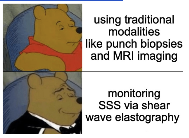
What are the clinical and radiological features of stiff skin syndrome, and can shear wave elastography change how we monitor disease progression?
Pediatric Dermatology
Pediatric Dermatology
Why did the skin specimen go to the party? It wanted to show off its "epiderm-dress" and have a "derma-blast!"
Stiff skin syndrome (SSS) is a rare disorder characterized by extremely hard and indurated skin, primarily caused by pathogenic mutations in the FBN1 gene with autosomal dominant inheritance. SSS presents in childhood or early adolescence with symmetric, sclerotic skin changes affecting various body parts, leading to functional limitations and frequent misdiagnosis.
This single-center cohort study contributes to the limited literature on SSS by delineating clinical and radiological findings of SSS in 11 pediatric patients (under age 18), along with exploring the efficacy of shear wave elastography (SWE) for surveillance of sclerotic skin changes.
What did they find?
Limitations: This study is limited by its retrospective component, small sample size, and lack of standardized controls for the prospective imaging studies.
Main Takeaway: SSS is characterized by segmental sclerotic changes, particularly in the pelvic and shoulder girdle regions, and SWE is a noninvasive and objective tool for evaluating and monitoring sclerotic changes.
Stiff skin syndrome (SSS) is a rare disorder characterized by extremely hard and indurated skin, primarily caused by pathogenic mutations in the FBN1 gene with autosomal dominant inheritance. SSS presents in childhood or early adolescence with symmetric, sclerotic skin changes affecting various body parts, leading to functional limitations and frequent misdiagnosis.
This single-center cohort study contributes to the limited literature on SSS by delineating clinical and radiological findings of SSS in 11 pediatric patients (under age 18), along with exploring the efficacy of shear wave elastography (SWE) for surveillance of sclerotic skin changes.
What did they find?
- This study found a predominance of SSS in males (54%, 6/11), with a median age of disease onset at 2.7 years (IQR 0-8)
- The cutaneous presentation of SSS is mostly segmental (90%, 10/11), with unilateral (64%, 7/11) or bilateral (36%, 4/11) skin changes characterized by thickened, bound-down skin with a "rippling" appearance
- Clinical and radiological assessments revealed variable degrees of sclerotic changes (36%, 4/11), hypertrichosis (18%, 2/11), lipodystrophy (18%, 2/11), limited range of motion (18%, 2/11), and functional limitations (18%, 2/11)
- Prospective MRI imaging studies showed abnormal high signal intensity in affected tissues correlating with anatomical sites involved, predominantly in the shoulder and pelvic girdle
- Shear wave elastography (SWE) consistently showed higher values within the dermis of the pathological site (32.8 kPa, IQR 18.5-43.0 kPa) compared to the control site (18.3 kPa, IQR 14.9-20.9), indicating an appropriate increase in dermal stiffness over repeated evaluations
Limitations: This study is limited by its retrospective component, small sample size, and lack of standardized controls for the prospective imaging studies.
Main Takeaway: SSS is characterized by segmental sclerotic changes, particularly in the pelvic and shoulder girdle regions, and SWE is a noninvasive and objective tool for evaluating and monitoring sclerotic changes.
Does dupilumab improve sleep in adults with atopic dermatitis?
British Journal of Dermatology
British Journal of Dermatology
Catch some zzz’s with dupilumab!
Atopic dermatitis (AD) is characterized by extremely itchy eczematous plaques; the itch results in sleep disturbance, causing day-time fatigue and impacting quality of life. Dupilumab is a monoclonal antibody that inhibits IL-4 and IL-13 signaling and improves inflammation in AD.
In this study, the authors sought to explore whether dupilumab improved the sleep disturbance in AD through measuring both sleep quality and duration. They examined 188 adults with moderate-to-severe AD after 12 weeks of dupilumab or placebo.
What did they find?
Limitations: The primary endpoint in this study relied on sleep NRS scoring, a relatively new rating scale. As such, there are not many validating data to support the use of sleep NRS. However, the authors also saw similar improvement in sleep disturbance using other sleep rating scales. Furthermore, this study only evaluated one time point (12 weeks). Longer studies are required to see how long dupilumab’s effects on sleep may persist for.
Main takeaways: Dupilumab improved sleep disturbance in adults with AD after 12 weeks of treatment.
Atopic dermatitis (AD) is characterized by extremely itchy eczematous plaques; the itch results in sleep disturbance, causing day-time fatigue and impacting quality of life. Dupilumab is a monoclonal antibody that inhibits IL-4 and IL-13 signaling and improves inflammation in AD.
In this study, the authors sought to explore whether dupilumab improved the sleep disturbance in AD through measuring both sleep quality and duration. They examined 188 adults with moderate-to-severe AD after 12 weeks of dupilumab or placebo.
What did they find?
- Dupilumab resulted in clinically meaningful reduction in itch
- Sleep quality significantly improved in patients on dupilumab
- The proportion of patients reporting treatment-related adverse effects was lower in those receiving dupilumab than those receiving the placebo
Limitations: The primary endpoint in this study relied on sleep NRS scoring, a relatively new rating scale. As such, there are not many validating data to support the use of sleep NRS. However, the authors also saw similar improvement in sleep disturbance using other sleep rating scales. Furthermore, this study only evaluated one time point (12 weeks). Longer studies are required to see how long dupilumab’s effects on sleep may persist for.
Main takeaways: Dupilumab improved sleep disturbance in adults with AD after 12 weeks of treatment.
Will Magnetic Resonance Imaging change the face of diagnosing eosinophilic fasciitis?
Journal of the American Academy of Dermatology
Journal of the American Academy of Dermatology
I’ll take the non invasive MRI please!
Skin biopsies are the current gold standard for diagnosis, providing physicians a cellular view of underlying pathology. Biopsies are, however, uncomfortable and invasive. Eosinophilic fasciitis (EF) is a skin condition classified by erythema, edema, and inflammation of the extremities in addition to sclerosis and thickening of the fascia that is typically diagnosed via wedge biopsy.
Physicians have utilized T2-weighted magnetic resonance imaging (MRI) for diagnosis and monitoring disease progression in patients with EF as it is noninvasive and allows for full evaluation of the extent of the disease. MRI may provide physicians with a noninvasive and less error-prone method of diagnosing EF.
What did they find?
Main Takeaway: MRI may provide physicians with a non-invasive diagnostic tool for patients with EF in place of wedge biopsy.
Skin biopsies are the current gold standard for diagnosis, providing physicians a cellular view of underlying pathology. Biopsies are, however, uncomfortable and invasive. Eosinophilic fasciitis (EF) is a skin condition classified by erythema, edema, and inflammation of the extremities in addition to sclerosis and thickening of the fascia that is typically diagnosed via wedge biopsy.
Physicians have utilized T2-weighted magnetic resonance imaging (MRI) for diagnosis and monitoring disease progression in patients with EF as it is noninvasive and allows for full evaluation of the extent of the disease. MRI may provide physicians with a noninvasive and less error-prone method of diagnosing EF.
What did they find?
- 68 patients were included in a study with 41 patients who underwent a full-thickness wedge biopsy, 46 who underwent MRI, and 23 who had both
- The sensitivity for detecting fascial changes consistent with EF using MRI was 93.5% while the sensitivity of detecting fascial changes in the wedge biopsy was 95.1%.
- 82.6% of the patients that underwent both modalities had EF detected on both MRI and wedge biopsy
- The difference in sensitivity had a P-value of 0.99, showing no statistical significance between the MRI and wedge biopsy for detecting EF fascial change
Main Takeaway: MRI may provide physicians with a non-invasive diagnostic tool for patients with EF in place of wedge biopsy.
Kikuchi-Fujimoto disease (KFD) is a rare, necrotizing lymphadenitis thought to be an immune-mediated response to infection that presents with a flu-like prodrome, fevers, and lymphadenopathy. Common cutaneous manifestations of KFD include erythematous macules, patches, plaques, malar erythema, pruritus, and oral ulcers. We discuss a rare case of KFD initially presenting with an acneiform eruption with concurrent systemic lupus erythematosus (SLE).
Report of Case:
Report of Case:
- A 48-year-old female with a history of Graves disease presented to the ED with several weeks of nausea, vomiting, intermittent fevers, fatigue, and a nonpruritic, nontender rash
- On exam, the patient had multiple erythematous-to-violaceous, follicularly-centered macules and papules with central crust on the preauricular cheeks, nasal dorsum, and forehead
- Our clinical evaluation was considered along with skin and lymph node biopsy findings and positive ANA and anti-smith serologies, and the patient was diagnosed with KFD and concurrent SLE


