fourth issue
february 23, 2022
Does spironolactone really increase your risk for cancer?
JAMA Dermatology
Spironolactone is taking the world by storm with many people crediting it for fixing their refractory acne, but is it all it’s cracked up to be? The off-label use of spironolactone for acne, hirsutism, and alopecia has increased in popularity and proven to be an effective alternative for acne treatment. However, the FDA has an official warning for increased tumorigenicity begging the question of whether dermatologists should continue touting it as an option for treatment. This systematic review and meta-analysis reviewed seven studies with a total population of 4,528,332 individuals. The results showed no statistically significant association between spironolactone use and risk of breast cancer (risk ratio [RR], 1.04; 95% CI, 0.86-1.22), ovarian cancer, bladder cancer, kidney cancer, gastric cancer, or esophageal cancer. There was an association between spironolactone use and decreased risk of prostate cancer (RR, 0.79; 95% CI, 0.68-0.90). The results are reassuring that spironolactone is unlikely to be associated with a substantial increased risk of cancer and prescribing could be considered. However, the study was limited by low certainty of evidence and need for inclusion of more diverse populations such as younger individuals and those with acne or hirsutism so stay tuned!
Selective phosphodiesterase 4 inhibitors: a new topical option for treating atopic dermatitis?
Journal of American Academy of Dermatology
Are you saying dermatologists can use something OTHER than topical steroids?! Atopic dermatitis (AD) is a very common, chronic, and pruritic inflammatory skin disorder. While topical steroids and topical calcineurin inhibitors (e.g. tacrolimus and pimecrolimus) have been mainstays in treatment, there have been concerns over their safety profile with long-term usage. For this reason, new topical therapies have been developed, including selective phosphodiesterase 4 (PDE4) inhibitors (i.e. difamilast), which target a key player in the pathogenesis of AD. In this phase 3 randomized, double-blind, vehicle- controlled clinical trial, efficacy and safety was compared in an adult Japanese population with atopic dermatitis who received difamalist ointment 1% (n=182) versus a vehicle (n=182) twice daily for 4 weeks. The percentage of patients achieving an investigator global assessment score of 0 or 1 with 2+ grade improvement at week 4 (primary endpoint) was significantly higher in the difamilast-treated population than vehicle (38.46% vs 12.64%, respectively, P < .0001). Regarding safety, 32 patients in the 1% difamilast group and 51 patients in the vehicle group experienced treatment-emergent adverse events, although most were mild and included worsening of AD followed by nasopharyngitis. Limitations to this study include that it only included Japanese patients, which may reduce its generalizability. Future studies could focus on longer-term evaluations and directly compare selective PDE4 inhibitors to topical calcineurin inhibitors and steroids.
Getting Rid of the Itch with Ruxolitinib
Journal of Investigative Dermatology
Ever had that itch you can’t scratch? Well, those living with lichen planus literally may experience that every day. Lichen planus (LP) is a chronic inflammatory condition of the skin and mucous membranes causing demarcated pruritic papules and plaques; approximately 1 in every 100 people have LP. The pathogenesis of LP occurs through an interferon-gamma (IFN‐γ) mediated JAK2/STAT1 pathway. Ruxolitinib, a JAK1/2 inhibitor which targets IFN‐γ, may help patients with LP. Researchers at Mayo Clinic enrolled 12 participants with LP in a prospective, open-label study, to apply ruxolitinib twice per day for 8 weeks. Most patients included in the study did not respond well to other systemic therapies. Patient-reported quality of life increased significantly throughout the treatment, providing evidence that JAK 1 and/or 2 play a critical role in pruritis in LP. The total number of lesions decreased by a median of 50 lesions (p<0.001), and lesion severity scores decreased by a mean difference of 7.6 points on the mCAILS scale (p=0.016). This study is limited by its small sample size and lack of placebo-control. However, preliminary findings are promising that topical ruxolitinib can be helpful in the treatment of LP.
QUESTION OF THE WEEK
NEJM CHALLENGE QUESTION
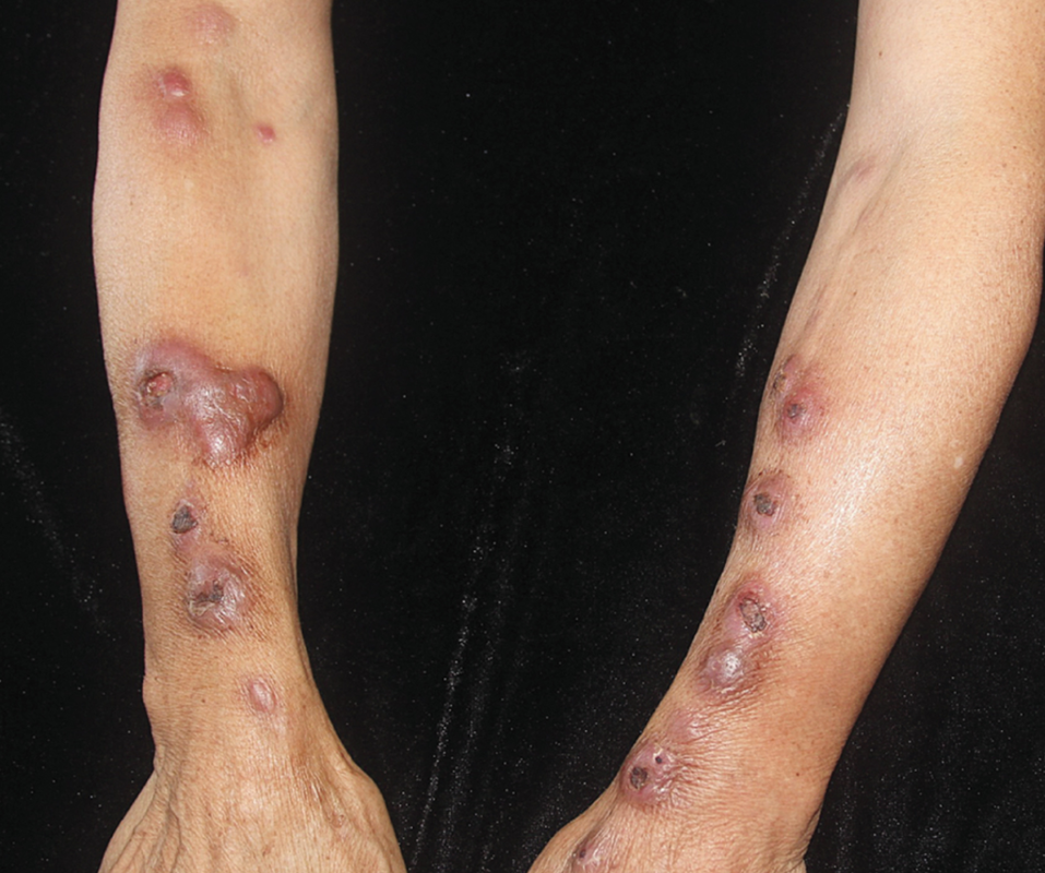
A 63-year-old woman presented with an 8-week history of nodules on her arms. A biopsy was performed and demonstrated multiple granulomas. What is the diagnosis?
- Squamous cell carcinoma
- Mycobacterium marinum
- Syphilis
- Sarcoidosis
- Herpes simplex virus infection
Answer:
Mycobacterium marinum. This bacterium is found in aquatic environments, including fresh, salt, and brackish water. This woman gave a history of washing fish with a cut on her thumb 6 weeks before the nodules developed.
Mycobacterium marinum is caused by skin to skin contact (usually through abrasions on the hand) with aquatic environments, such as fish tanks. The clinical presentation is erythematous to blue ulcerating nodules in a “sporotrichoid pattern”. In our case, this was misdiagnosed as sporotrichosis originally. The diagnosis is confirmed with culture and treatment is with clarithromycin and rifampin/ethambutol, minocycline and bactrim may be added on. Of note, M. marinum grows best at 31 degrees Celsius whereas other mycobacteria grow at 37 degrees.
Other answers:
A. Squamous-cell carcinoma - This typically presents as an erythematous scaly papulonodule or plaque on the head/neck and dorsal extremities (most common locations). Risk factors include chronic sun exposure, male gender, older age, fair skin types, immunosuppression, HPV, chronic non-healing wound, hypertrophic lichen planus or lupus erythematosus, chronic lichen sclerosis et atrophicus, and arsenic exposure. Other risk factors include patients with CLL, undergoing treatment with vemurafenib (BRAF inhibitor), long-term voriconazole prophylaxis and organ transplant (65x).
C. Syphilis- Secondary syphilis can have various skin manifestations and typically presents 3-10 weeks after the painless chancre (primary syphilis). Prodromal signs include malaise, fever, lymph node enlargement. Cutaneous manifestations include a papulosquamous/maculopapular generalized rash that is “copper-colored” with papules and plaques on the palms and soles. You may also see “moth eaten alopecia”, mucous patches in oropharynx, hypopigmented macules on neck (“necklace of venus”), and condyloma lata.
D. Sarcoidosis- This entity has a bimodal peak at 25-30 years old and 45-65 years old and has higher incidence in females and African American populations. Sarcoidosis classically presents as red-brown or erythematous papules and plaques with characteristic “apple jelly” color with diascopy. Lesions typically do not have secondary change and they occur most frequently on the face (especially lips and nose), neck and upper half of body.
E. Herpes simplex virus infection- This appears as grouped or clustered vesicles on an erythematous base with orolabial lesions being caused typically by HSV1 and genital lesions caused by HSV2 (classically).
